Figure 3.

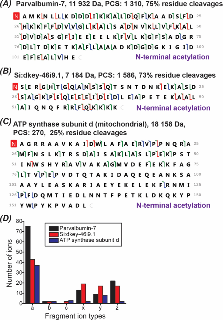
Proteoform fragmentation data. (A)-(C): sequences and fragmentation patterns of Parvalbumin-7, Si:dkey-46i9.1, and ATP synthase subunit d (mitochondrial). a/x ions, b/y ions, and c/z ions were marked in green, blue, and red. (D): Distribution of the fragment ion types for the three proteoforms shown in (A)-(C).
