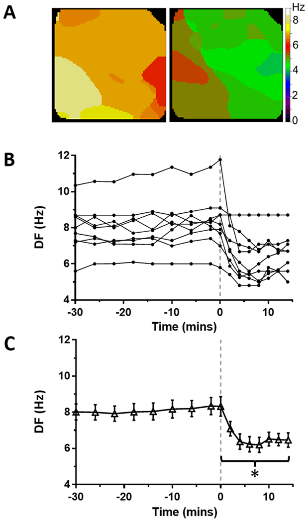
Figure 4: Effect of ranolazine on DF in tachypacing-induced persistent AF on the LA epicardium from perfused hearts.

(A) Time course of maximal dominant frequency (DF) during persistent AF in 9 sheep hearts. Continuous ranolazine perfusion (10 μM) begins at time 0 (dashed line). (B) Average DF was decreased during ranolazine perfusion (*p<0.01). (C) Representative DF maps of the LA epicardium before (left) and during (right) perfusion of ranolazine.
