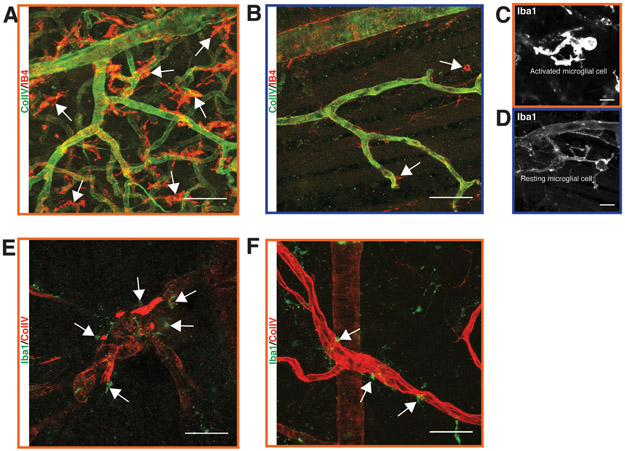
Figure 8.
Microglial infiltration during diabetic retinopathy in the Nile rat. A: Abnormally high density of microglial cells in a retina with proliferative diabetic retinopathy. B: An age-matched control of panel A. C: An activated microglial cell at high magnification from the same retina as in panel A. D: A resting microglial cell at high magnification from the same retina as in panel B. E: Several microglial cells surround a highly tortuous arteriolar structure in a diabetic retina. F: A few microglial cells surround a dilated venule in a diabetic retina. Scale bars of A, B, E and F = 50 μm. Scale bar of C and D = 10 μm.

