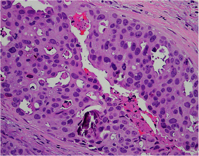
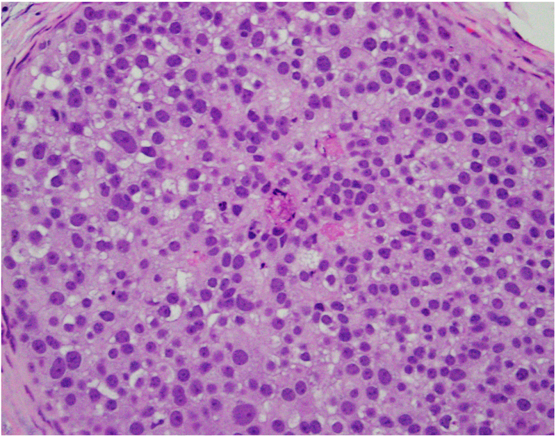
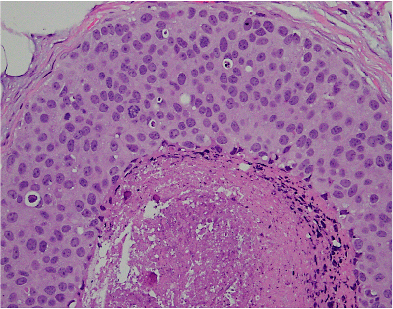
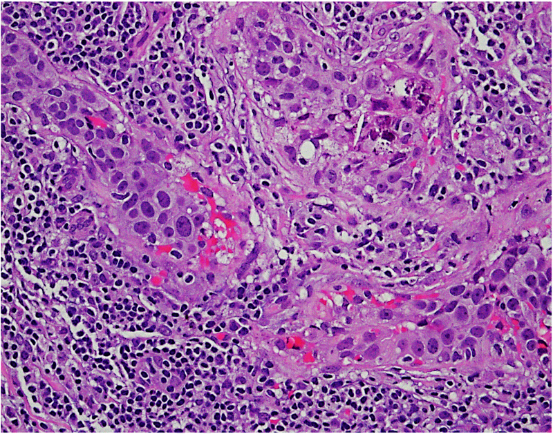
Fig. 2.
(A–D) Heterogeneity in the morphological patterns of ductal carcinoma in situ (DCIS). Images are taken from a needle biopsy of a single patient with DCIS (same patient as Fig. 1; 20× objective using a Nikon DP27 camera using CellSens software). Note the range of architectural patterns ranging from solid to cribriform as well as the variable amounts of necrosis in the ducts.


