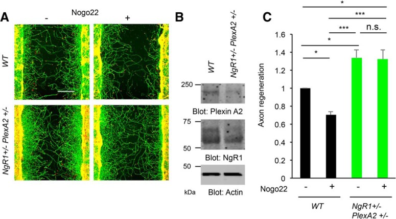
Figure 6.
NgR1+/−PlexinA2+/− neuron shows enhanced axonal regeneration in vitro. A, Cortical neurons from WT and NgR1+/−PlexinA2+/− were scraped at 8 DIV and allowed to regenerate for 3 d in the presence of Nogo22 (100 nm). The microphotographs show βIII tubulin (in axons; green) and phalloidin (to stain F-actin; red) to illustrate the growth cones of cortical neurons in the middle of the scraped area. Scale bar, 200 μm. B, Immunoblots of lysates cultures with anti-NgR1, PlexinA2, and actin antibodies. C, The graph shows quantification of axonal regeneration normalized to WT control (Nogo22-). Error bars represent SEM; n = 5 biological replicates from different embryos. n.s., not significant, *p < 0.05, ***p < 0.0005, one-way ANOVA followed by Tukey's test.

