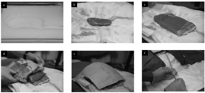
Fig. 1.
Ex-vivo partial pig model setup. (a) Foam container with cut-out kidney impression. (b) Pig kidney with ureter placed on absorbent pad on foam. (c) Pig flank placed overtop of kidney. (d) Further pig flank slabs, including ribs placed overtop to simulate human anatomy. (e) Complete setup of partial pig model: final layer consists of skin and subcutaneous tissue. (f) 5 Fr open-ended ureteric catheter is placed up ureter and tied in place, contrast syringe is attached to external end of catheter.

