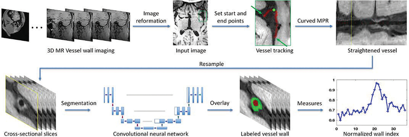
Fig. 1.
Image processing pipeline proposed in IVA framework. Top row shows the image preparing steps and bottom row demonstrates the image segmentation and quantification steps. The originally acquired 3D VWI image set is first reviewed to identify the interested intracranial vessel (the green box). Then this region is zoomed up where the start and end points of the vessel could be manually designated. It is followed by vessel centerline tracking and vessel straighten using curved MPR (the yellow line in the “Straightened vessel” panel corresponds to the location marked by a greed dot in the “vessel tracking” panel), and sliced into contiguous cross-sectional 2D slices that will in turn undergo vessel segmentation and quantification analysis.

