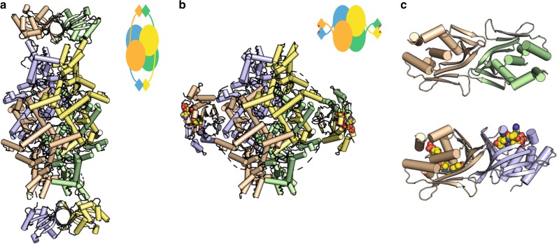
Fig. 1.
Structure of ICL2. a Ligand-free ICL2 forms an elongated structure with C-terminal dimers at each end of the structure. b Striking structural rearrangement of ICL2 upon binding to acetyl-CoA. In both a and b, each monomer is shown in different colours and the schematics outline the structural features of the tetramer in each case. c The dimeric association of the C-terminal domains in the ligand-free (top) and acetyl-CoA-bound (bottom) ICL2. Acetyl-CoA is shown as spheres in panels b and c. Both panels are shown with the same orientation of the wheat-coloured monomer

