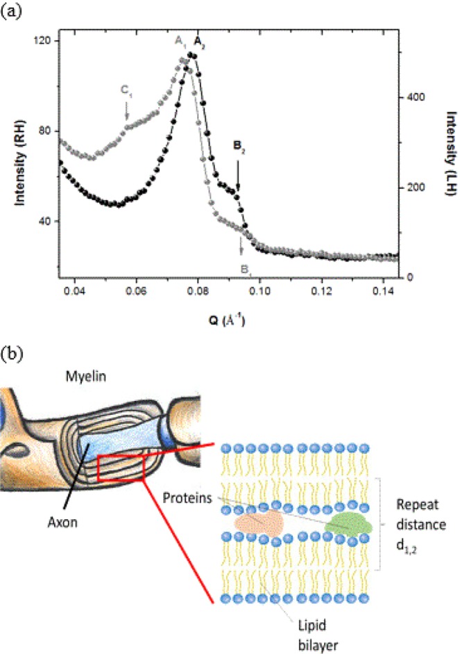
Figure 1.

Panel a: Diffraction patterns obtained for the RH (black symbols) and LH (gray symbols) at 300 K measured on D16 at ILL. Second order Bragg peaks are found at A1 = 0.078 Å−1 (RH) and A2 = 0.074 Å−1 (LH). B1 and B2 represent less pronounced first (1st) or second (2nd) order Bragg peaks at Q = 0.093 Å−1 (RH) and Q = 0.088 Å−1 (LH). Additional reflection (C1) is observed in LH at Q = 0.057 Å−1. Panel b: Sketch of a myelin sheath and extraction of the multilamellar lipidic structure with attribution of the repeat distance d1 or d2.
