Fig. 3.
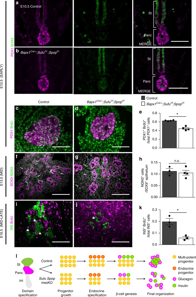
Mesenchymal loss of Sufu and Spop impairs progenitor growth and beta cell genesis. a, b Immunostaining for the SOX2+ stomach domain (St) and PDX1+ pancreatic domain (Panc) in E10.5 control a and Bapx1Cre/+;Sufuf/f;Spopf/f mutants b embryos. c, d Immunostaining for E10.5 PDX1+ control c vs. mutant d pancreatic progenitors marked with thymidine analog, 5-bromo-2′-deoxyuridine (BrdU) 1 h after administration. e Assessment of the proportion of PDX1+ progenitors marked by BrdU as a measure of progenitor proliferation in mutants vs. controls (n = 4 samples per genotype). f, g Immunostaining for E13.5 NGN3+ control f vs. mutant g endocrine progenitors in the SOX9+ pancreatic progenitor pool. Arrows indicate non-specific staining. h Quantification of the proportion of NGN3+ committed endocrine progenitors out of the total NGN3+ and/or SOX9+ progenitor pool in mutants vs. controls (n = 3 control samples, n = 4 mutant samples). i, j Immunostaining for control i vs. mutant j INS+ cells labelled with BrdU after administration every 24 h during beta cell genesis (E14.5-E16.5). Arrows indicate representative BrdU+ labelled INS+ cells. k Assessment of INS+ cells labelled with BrdU after E14.5-E16.5 tracing as a measure of beta cell genesis in mutants vs. controls (n = 3 samples per genotype). l Diagram illustrating the temporal roles of mesenchymal Sufu and Spop throughout pancreatic development. Data are means ± SEM. n.s. denotes not significant, * denotes p < 0.05 by Student’s un-paired, two tailed t-test. Scale bars- 100 µm

