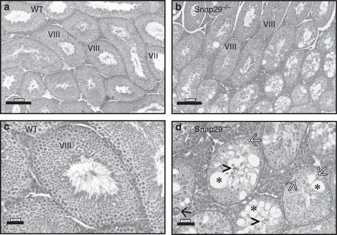
Fig. 8.
Histological analysis of Snap29−/− testis. Testis sections were stained with hematoxylin and eosin to evaluate spermatogenesis in WT (a, c) and Snap29−/− (b, d) mice. WT testis displayed normal spermatogenesis (Stages VII and VIII showing elongating spermatids and spermatozoa in the lumen). Seminiferous tubules in Snap29−/− testis had degenerated germ cells (white arrows), loss of immature germ cells accumulated in the lumen (arrow heads), giant multinucleated spermatids (black arrow) and extensive vacuolization (*). The diameter of degenerated seminiferous tubules was reduced in Snap29−/− (d) compared with WT (c) testis

