Figure 6.
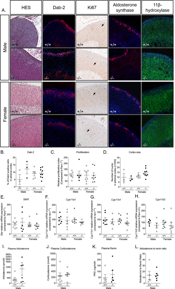
Adrenal cortex disorganization persists with aging in Rarα−/− mice. (A) Morphological characterization of adrenals from 52 weeks old male and female Rarα+/+ and Rarα−/− mice. HES staining, Dab-2, aldosterone synthase and 11β-hydroxylase immunofluorescence and Ki67 immunohistochemistry were performed. (B) Number of Dab-2 positive cells in the cortex was determined in 3 to 9 animals of each genotype and sex using an automated molecular imaging platform (Vectra, Perkin Elmer) and is expressed as a percentage of total number of cells in the entire cortex area. (C) Relative proliferative index of adrenals from male and female Rarα+/+ and Rarα−/− mice. Ki67 positive cells were separately counted in the adrenal cortex in 5–6 animals per genotype. (D) Number of nuclei in the adrenal cortex was determined in 3 to 9 animals of each genotype and sex using an automated molecular imaging platform (Vectra, Perkin Elmer). (E–H) Expression of steroidogenic genes in male and female Rarα+/+ and Rarα−/− mice. mRNA expression of Star (E), Cyp11a1 (F), Cyp11b1 (G) and Cyp11b2 (H) was assessed by RT-qPCR. RT-qPCR were performed on mRNA extracted from 6–8 adrenals from 52 weeks old male and female Rarα+/+ and Rarα−/− mice. (I,J) Measure of plasma aldosterone (I) and corticosterone (J) concentration by mass spectrometry in male mice. (K,L) Plasma renin concentration (PRC) and aldosterone to renin ratio. Measure of plasma aldosterone, plasma corticosterone and plasma renin were done on 5–6 animals per group. Values are presented as means ± SEM. *p < 0.05; **p < 0.01.

