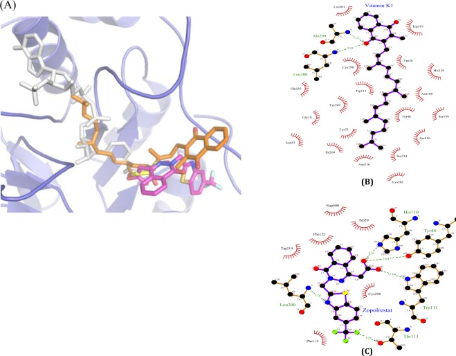
Figure 7.
(A) Close-up view of docked conformation of vitamin K1 in ALR2 shows the overlap with the NADPH (white sticks, PDB id: 4IGS), Zopolrestat (magenta sticks, PDB id: 2HVO), and glyceraldehyde (yellow sticks, PDB id: 3V36) binding sites. 2D representation showing the interactions of vitamin K1 (B) and Zopolrestat (C) with ALR2 residues. The ligands and protein are shown with purple and brown bonds respectively. Hydrogen bond are shown in dotted lines along with donor-acceptor distance and residues interacting by hydrophobic interactions were represented as lines in red. The diagrams are prepared using LIGPLOT (Wallace et al., (1996). Protein Eng., 8, 127–134).

