
Figure 3.
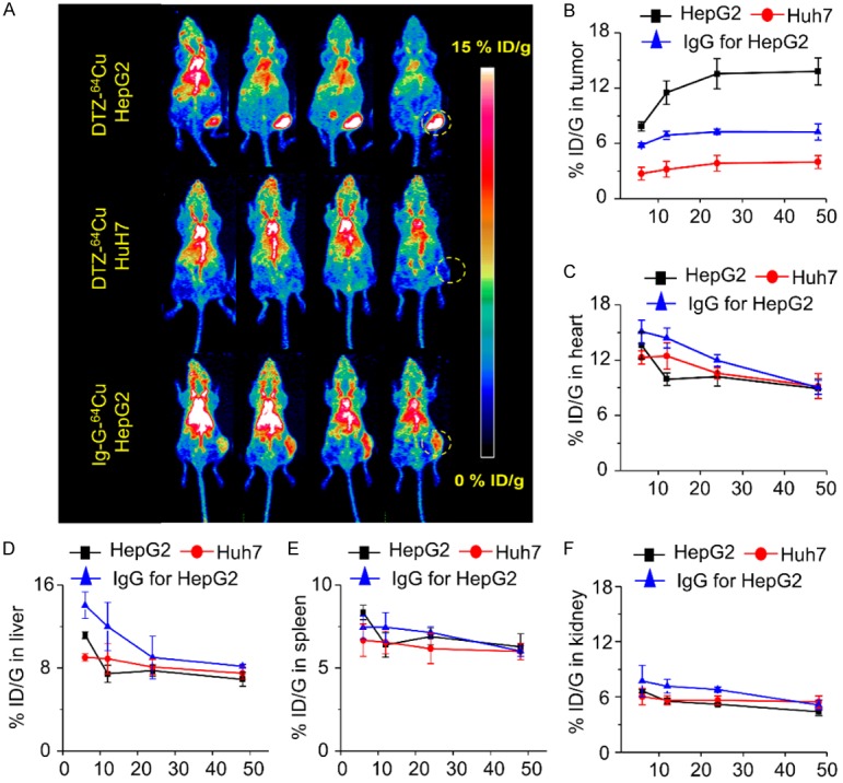
(A) PET maximum intensity projection (MIP) images of HepG2 and Huh7 lymphoma tumor-bearing models from 6 to 48 h post-injection of 64Cu-NOTA-daratumumab (DTZ-64Cu). Quantitative results of PET imaging in tumor (B), heart (C), liver (D), spleen (E) and kidney (F) after injection of 64Cu-NOTA-daratumumab or 64Cu-NOTA-IgG in HepG2 and Huh7 liver tumor models. n=4 per group.