Figure 2.
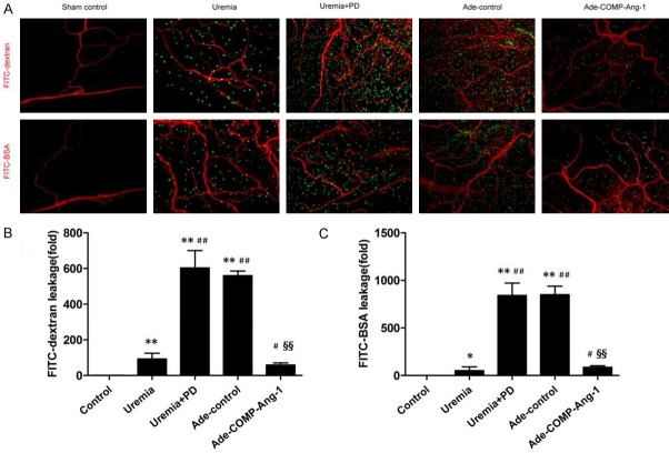
COMP-Ang-1 ameliorates uremia and bioincompatible PD fluid-induced peritoneal microvascular hyperpermeability. After PD for 4 weeks, rats were anesthetized with 9% chloral hydrate. FITC-BSA (69 kDa, 10 mg/100 g, 2 ml/100 g, Sigma-Aldrich, St. Louis, MO) or FITC-dextran (4 kDa, 10 mg/100 g, 2 ml/100 g, Sigma-Aldrich, St. Louis, MO) was injected via the tail vein. Systemic blood vessels were lavaged with saline to wash out intravascular fluorescent substances after 20 min. After rats were euthanized, mesenteric samples were separated and trimmed to create a flat surface and stained with the immunofluorescent antibody against CD31 (a marker of endothelial cells). A. Immunofluorescence staining for FITC-dextran and FITC-BSA in visceral peritoneal membranes (magnification, × 100). Red indicates the endothelial cell marker CD31. Green indicates FITC-conjugated dextran or BSA. B, C. Semiquantitative analysis of peritoneal vessel permeability to FITC-dextran and FITC-BSA in the control, uremia nondialysis, uremia+PD, Ade-control and Ade-COMP-Ang-1 groups. Data are presented as the means ± SDs (n = 6 per group). *P < 0.05 versus the control group; **P < 0.01 versus the control group; #P < 0.05 versus the uremia group; ##P < 0.01 versus the uremia group; §§P < 0.01 versus the uremia+PD group.

