Figure 3.

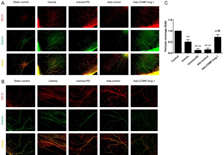
COMP-Ang-1 preserves peritoneal pericyte attachment after uremia and bioincompatible PD fluid exposure. After being processed as described in the Materials and Methods, mesenteric samples were stained with immunofluorescent antibodies against CD31 (a marker of endothelial cells, red) and Desmin (a specific marker of pericytes, green). A, B. CD31/Desmin double immunofluorescence staining in visceral peritoneal membrane sections (magnification, × 40 and × 100, respectively). Red indicates the endothelial cell marker CD31. Green indicates the pericyte marker Desmin. C. Semiquantitative analysis of pericyte coverage measured in visceral peritoneal membrane sections from six rats in each group. Data are presented as the means ± SDs. **P < 0.01 versus the control group; ##P < 0.01 versus the uremia group; §§P < 0.01 versus the uremia+PD group.
