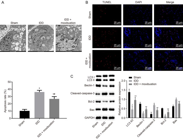
Figure 4.
Treatment with moxibustion restrains apoptosis and induces autophagy in NP cells. A. Micrographs showing autophagy of NP cells observed under transmission electron microscope. B. Apoptosis of NP cells detected by TUNEL staining (scale bar = 25 µm). C. The protein expression of autophagy and apoptosis-related genes LC3 II/I, Beclin-1, cleaved-caspase-3, Bcl-2 and Bax normalized to GAPDH in NP cells measured using western blot analysis. *P < 0.05 vs. sham-operated rats; #P < 0.05 vs. IDD rats. The measurement data were expressed as the mean ± standard error. Comparisons among multiple groups were analyzed using one-way ANOVA (Tukey’s post hoc test). Three parallel experiments were repeated.

