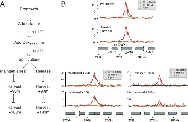
Fig. 2.
Histone tracking through DNA replication. (A) Schematic of experimental design. Before the experiment, cells were grown without reaching saturation for at least 8 doublings. (B) For each time and condition, a single sample was harvested, and the purified chromatin was divided and used to precipitate TetR-BirA(s/213) (anti-V5) and biotinylated histone H3-Avi (streptavidin), then sequenced to measure enrichment. Biotinylated histone density did not change in shape, indicating that histones did not move locally along the chromatin fiber; however, the total amount of biotinylated H3-Avi detected (rel. background biotinylation) decreased during an extended G1 arrest or during cell division. Results were representative of 2 biological replicates.

