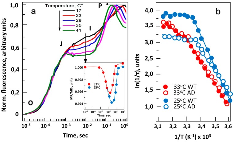
Figure 2.
The effect of temperature on chlorophyll fluorescence induction in Synechocystis. (a) Wild-type (WT) strain. Characteristic induction curves obtained at 17, 23, 29, 35 and 41 °C; letters O, J, I and P indicate different stages of plastoquinone (PQ) reduction, according to standard nomenclature. Insert panel: the effect of temperature on kinetics of modulated reflection (MR) at 820 nm obtained at 25 and 32 °C. (b) Arrhenius plots of P700+ reduction rates of wild-type strain (WT, solid symbols) and desA−/desD− mutant (AD; open symbols) at 33 °C (red circles) and 25 °C (blue circles). Cell cultures were dark-adapted for 5 min prior to MR measurements. MR changes were induced by array of red (627 ± 10 nm) light-emitting diodes (LEDs) delivering 3000 µmol photons/m2 sec of actinic light to a sample [59].

