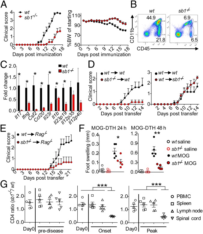
Fig. 2.
CD4 cell autonomous deficiency of Sb1 ameliorates EAE. (A–C) Wt and Sb1−/− mice were immunized with MOG/CFA to induce EAE. (A) Mean clinical score (Left) and body weight (Right) of wt (n = 13) and Sb1−/− (n = 14) mice. Experiment was repeated more than 5 times with the same pattern. (B) Spinal cord infiltrates on day 10 analyzed by FACS. n = 4–5 mice in each genotype, shown are representative of five experiments. (C) Relative gene expression of spinal infiltrates analyzed by qRT-PCR. Data represent mean of four biological replicates, each with pooled cells from 2 to 3 mice per genotype. (D) Adoptive transfer EAE. Wt or Sb1−/− T cells from MOG-immunized mice were expanded ex vivo and transferred to naive wt or Sb1−/− recipients. Mean clinical scores for 6 mice each genotype. (E) Naive CD4 cell transfer EAE. Wt or Sb1−/− naïve CD4 cells were transferred to Rag1/− mice, which were then MOG-immunized to induce EAE. Mean clinical scores for 6 mice each genotype. (F) DTH response of wt and Sb1−/− mice to challenge in the footpad with MOG or vehicle on day 6 after MOG immunization. (G) Ratio of Sb1−/− to wt CD4 cells in active EAE in chimeric mice. Symbols indicate individual mice. Data are representative of two (D, F, and G) or three (E) experiments. Error bars indicate ±SEM, *P < 0.05; **P < 0.01 by Student’s t test (C and F); ***P < 0.001 by one-way ANOVA (G).

