Fig. 6.
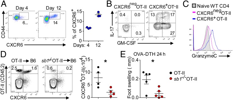
CXCR6 expression on OT-II cells. (A–C) OT-II cell transfer studies. Naïve OT-II cells (CD45.2) were transferred into naïve congenic CD45.1 mice, and the mice were immunized with OVA/CFA. (A) CXCR6 expression on LN OT-II cells on days 4 and 12 after immunization. (Left) Representative contour plots. (Right) Cell frequencies. (B) Cytokine profile of CXCR6neg and CXCR6+ OT-II cells on day 12 analyzed by FACS. (C) Histogram of GzmC expression. (D and E) Sb1−/− OT-II transfer studies. Naïve wt OT-II and Sb1−/− OT-II cells were separately transferred as in A, and the mice were immunized with OVA/CFA. (D) CXCR6-expressing wt and Sb1−/− OT-II cells in LN on day 10. (Left) Representative contour plots. (Right) Mean number of cells. (E) OVA-induced DTH (footpad swelling) in mice transferred with wt or Sb1−/− OT-II cells and challenged in the footpad. Symbols represent individual mice. Data are representative of three (A) and two (B–E) experiments. *P < 0.05 by Student’s t test.

