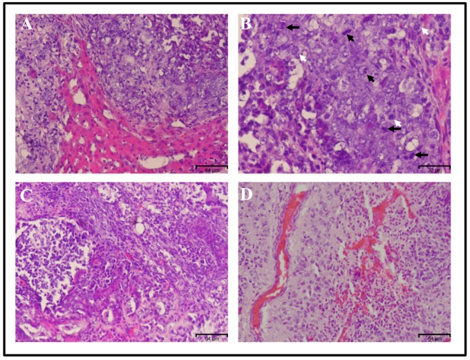
Figure 4.
Histological analysis of the tumor derived from transplantation of miPS-PLCcm cells. Hematoxylin and eosin staining showed tumor invasion inside the normal liver (A). Original magnification 20×. This tumor tissue showed malignant phenotype criteria such as mitotic figures (black arrows), nuclear atypia (white arrows) (B) Original magnification 40×, high nuclear to cytoplasmic ratio (C) and angiogenesis (D). Original magnification 20×.

