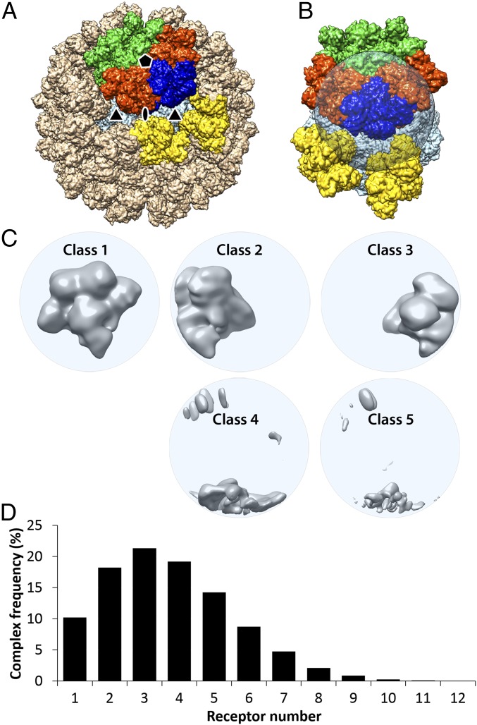
Fig. 3.
Focused 3D classification with symmetry-expanded orientation data characterizing the receptor binding sites based on the degree of receptor occupancy. (A) A simulation of 60 TfR:Tf heteromers bound to the CPV capsid was calculated by applying icosahedral symmetry operators to the bound TfR molecule in the symmetry-mismatch map (Fig. 2). The targeted volume occupied by the receptor was colored in blue; the 2 adjacent heteromers around the 5-fold axis were colored in orange; those across the symmetry axis were colored in green; those around the 3-fold symmetry axis were colored in yellow; and all other heteromers were colored in tan. (B) The transparent spherical mask applied for the target volume with occupying receptor colored in blue. (C) Five 3D-class reconstructions are shown in gray and surface rendered. The first class had full receptor densities. Classes 2 and 3 had partial densities corresponding to an unoccupied target volume plus the nearby orange receptors in A and B. The other classes had only some overlapping densities as shown. (D) A histogram for number of heteromers bound on each capsid. The average number of receptors bound to each capsid was 3.7.

