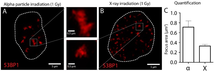
Figure 5.
Nanoscopic analysis of DSBs in U2OS cells. U2OS are irradiated using external alpha particle irradiation (A) or X-ray (B), fixed after 1 h and stained for 53BP1 as DSB marker. SIM imaging was used for nanoscopic analysis of 53BP1 foci. Foci were quantified using ImageJ (C). Enlarged figures show 53BP1 foci in close up.

