Figure 2.
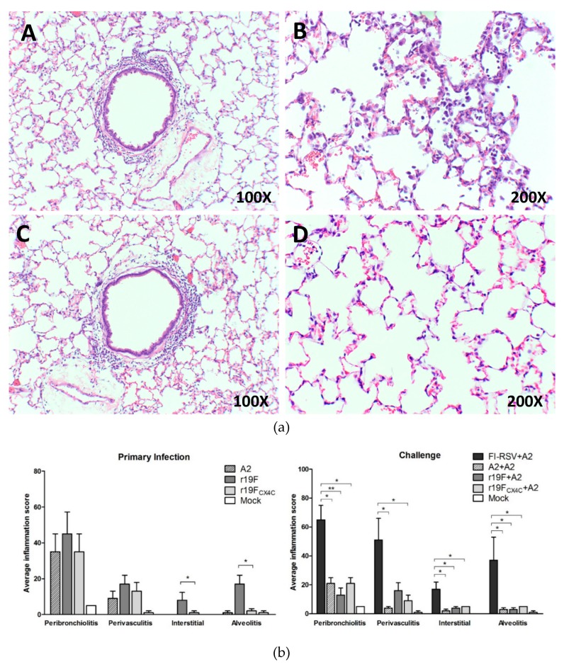
Lung histopathology 5 days after primary infection or challenge of cotton rats. Five female and/or male cotton rats (6–8 weeks) per group were infected intranasally with the 105 PFU of A2 or 2 × 10⁵ PFU of r19F or 2 × 10⁵ PFU of r19FCX4C in 0.1mL volume. Animals were sacrificed and lungs collected, fixed, and stained with hematoxylin and eosin. (a) Hematoxyline and eosin (H&E) staining of cotton rats lungs after primary infection with r19F (A,B) or r19FCX4C (C,D). Original magnifications are shown in the lower right corner of each panel. (b) Lung pathology scores after primary infection or A2 challenge. A total of four parameters were evaluated: peribronchiolitis (inflammatory cell infiltration around the bronchioles), perivasculitis (inflammatory cell infiltration around the small blood vessels), interstitial pneumonia (inflammatory cell infiltration and thickening of alveolar walls), and alveolitis (cells within the alveolar spaces). The slides were scored on a 0–4 scale and subsequently converted to a 0–100% histopathology scale. Note that no (0) interstitial (interstitial pneumonia) was seen on day 5 after primary infection with A2 or mock. Statistical significance is indicated: *, p < 0.05; **, p < 0.01 by nonparametric ANOVA (Kruskal–Wallis) and Mann–Whitney test.

