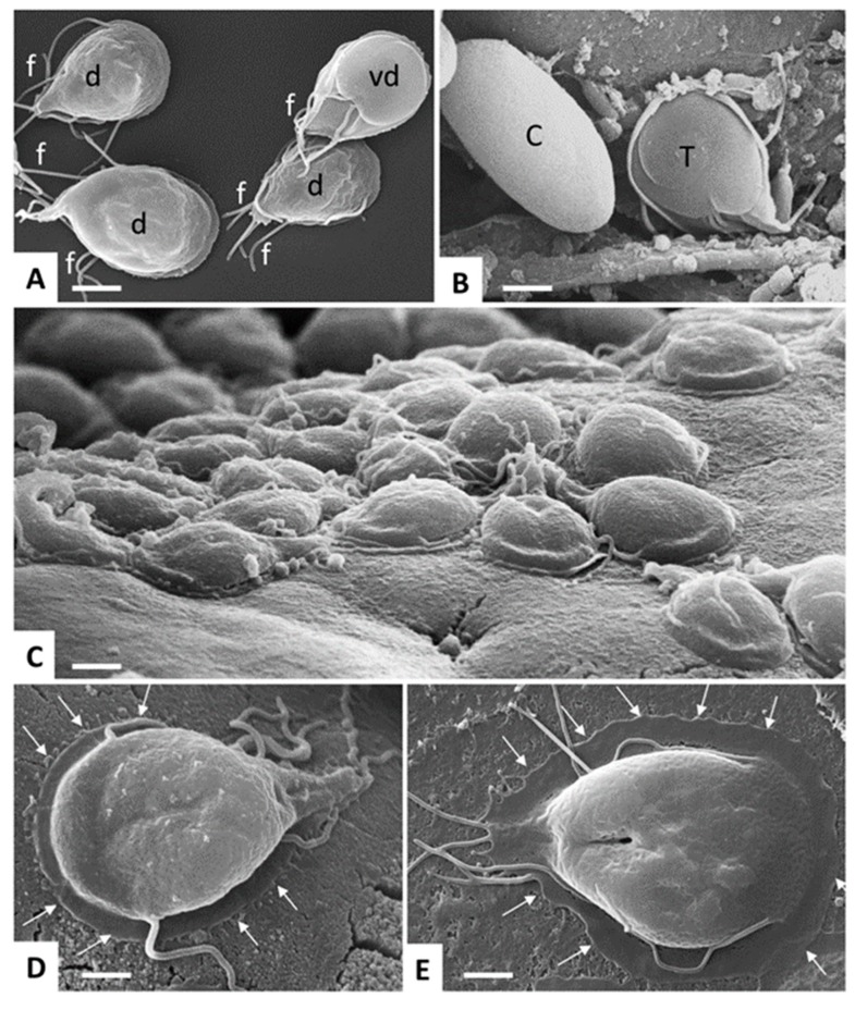
Figure 2.
Giardia lamblia trophozoites and cysts visualized by SEM. (A) trophozoites cultured in axenic in vitro culture, exposing either their dorsal surface (d) or ventral disc (vd). Note the multiple flagella (f). Bar = 8 µm. (B) Trophozoite (T) and cyst (C) stages in a mouse feces sample. Bar = 6.4 µm. (C) Trophozoites attaching to the intestinal surface of an experimentally infected mouse. Bar = 8 µm. (D,E) Higher magnification view of a trophozoite attaching to the mouse intestinal surface and to human colon carcinoma cells (Caco2) during in vitro culture, respectively. Arrows delineate the periphery of the ventral disc. Bar in (D,E) = 4 µm.

