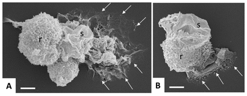
Figure 3.
Scanning electron microscopy of in vitro-cultured Entamoeba histolytica trophozoites. Note the different shapes and cell surface structures such as rough (r) and smooth (s) adopted by the trophozoites, and the cytoplasmic protrusions mediating contact to the surface (arrows). Bar in (A,B) = 5 µm.

