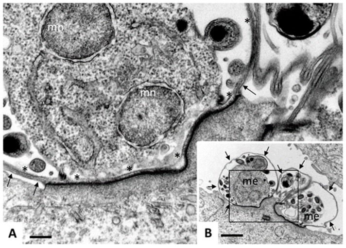
Figure 5.
Transmission electron microscopy (TEM) of Cryptosporidium parvum. Madin Darbey canine kidney (MDCK) cells were infected with C. parvum sporozoites and fixed and processed for TEM after 72 h of culture. Sporozoites have formed parasitophorous vacuoles (PVs) on the apical part of the MDCK cells, occupying a space which is still intracellular, but essentially extra-cytoplasmatic, giving rise to meronts. (B) A low magnification view of three PVs, with two developing meronts (me) clearly visible. Arrows indicate the outer host cell surface membrane. Bar = 12 µm. (C) A higher magnification view of the boxed area in (A). Asterisks (*) indicate the membrane of the parasitophorous vacuole, arrows point towards the host cell surface membrane, mn indicates nuclei of developing merozoites. Note the electron-dense zone where the PV is in close contact to the host cell cytoplasm, formed due to the cytoskeletal rearrangements. Bar = 2.5 µm.

