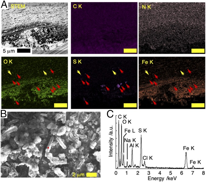
Fig. 3.
Cross-section and surface structure of white scaly-foot scale after 13-d incubation in the natural habitat of the black scaly-foot in the Kairei Field, CIR. (A) Dark-field STEM image of the thin section with EDS mapping taken at 25 kV; red and yellow arrows indicate sulfur-enriched area with and without iron, respectively. (Scale bars, 5 μm.) (B) SEM image of the surface of the translocated scale. (C) EDS spectrum of the mineral-coated bacteria indicated by a red dot in B.

