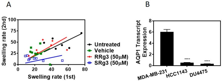
Figure 2.
(A) Double swelling assay showing the swelling rates for the first and the second swelling on a single oocyte. Eight oocytes per treatment were measured for swelling in a hypotonic medium, before and 2 h after exposure to a vehicle or epimers of Rg3. The results were analysed and presented with a linear regression. (B) The AQP1 transcript expression in MDA-MB-231, HCC1143 and DU4475 cell lines. Each data point represents a mean ± SD value of 3 replicates, and comparisons were made with the vehicle control group (**** p < 0.0001).

