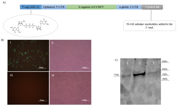
Figure 1.
(A) Genomic structure of naked conventional mRNA expressing the non-optimized S segment of the Ank-2 strain of CCHFV. To guarantee the stability of the structure, a 5′-cap, 3′-poly A tail, and 5′ and 3′-UTR were added. (B) The created mRNA vaccine construct was transfected in the BHK21-C13 cells and in vitro expression was verified by IIFA (×40): (i) fluorescence; (ii) phase contrast. We also included negative control cells as the background in the assay: (iii) fluorescence; (iv) phase contrast. (C) Expression of the His-tagged nucleocapsid protein in the bacterial system was analyzed by western blot assay. As became obvious after overnight IPTG induction of BL21 bacteria containing pET-N13, a strong band of 57 kDa (lane 2) was detected in the WB. As a control, we included the supernatant from the IPTG induced (lane 1) and the lysate from the non-induced bacteria containing the pET-N13 construct (lane 3).

