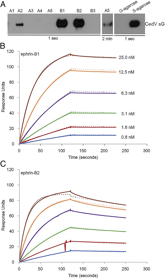
Fig. 1.
CedV G protein interacts strongly with both ephrin-B1 and -B2 and weakly with ephrin-A2 and -A5. (A) Coprecipitation of purified S-tagged CedV sG with a panel of soluble Fc-tagged ephrin molecules: human ephrin-A1, mouse ephrin-A2, human ephrin-A3, human ephrin A4, human ephrin-A5, mouse ephrin-B1, mouse ephrin-B2, and human ephrin-B3. CedV sG controls were precipitated with S protein agarose beads (S) or G protein agarose beads (G) in the absence of soluble ephrins. An overexposure of protein blot was included for coprecipitation with soluble ephrin-A5 and CedV sG. SPR (BIAcore) sensorgrams record the interaction in response units, between sG proteins and soluble (B) mouse ephrin-B1 or (C) mouse ephrin-B2. Cycles of receptor association and dissociation performed at 6 different receptor concentrations are shown. The continuous lines represent the experimental data, and the receptor concentrations are color-coded as indicated. A 1:1 Langmuir interaction model was used to fit the data (dotted line).

