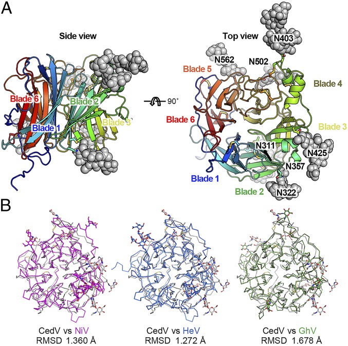
Fig. 4.
Structure of CedV G and a comparison with other henipavirus G proteins. (A) Top and side views of CedV G structures in cartoon diagrams. Disulfide bonds and N-linked glycans are shown as sticks and spheres, respectively. (B) Superimposition of head domain structures from selected henipaviruses.

