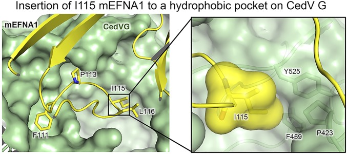
Fig. 8.
Structural basis for species specific usage of ephrin-A1. Structural modeling of residue T/I115 insertion into the CedV G receptor-binding pocket. CedV G is shown as green surface, while ephrin-A1 is shown in yellow. (Inset) I115 is shown as surface and sticks. Critical I115-contacting G protein residues are shown as sticks and labeled.

