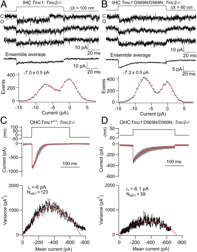
Fig. 6.
Single MET channel current amplitudes are unaltered in hair cells of Tmc1 p.D569N/D569N mice. (A) Single-channel records in the Tmc1+/+; Tmc2−/− mouse, ensemble average of 40 stimuli, and amplitude histogram with channel of −6.9 pA. (B) Single-channel records, ensemble average of 100 presentations, and amplitude histogram of Tmc1 p.D569N/D569N; Tmc2−/− with channel of −7.1 pA. A and B are for P6 mouse apical IHCs. (C) OHC MET currents in the Tmc1+/+; Tmc2−/− mouse with mean current (red) with individual responses (gray) for 50 stimuli superimposed. (Bottom) Plot of current variance σI2 versus current amplitude I, fit with parabolic equation (Materials and Methods) giving single-channel current io = −6.0 pA and number of channels NMET = 123. (D) OHC MET currents in Tmc1 p.D569N/D569N; Tmc2−/− mouse with mean current (red) with individual responses (gray) for 40 stimuli superimposed. (Bottom) Plot of current variance σI2 versus current I, gives io = −6.1 pA) and NMET = 59. C and D are for P6 mouse apical OHCs; extracellular Ca2+ was 0.04 mM (A and B) and 1.5 mM (C and D).

