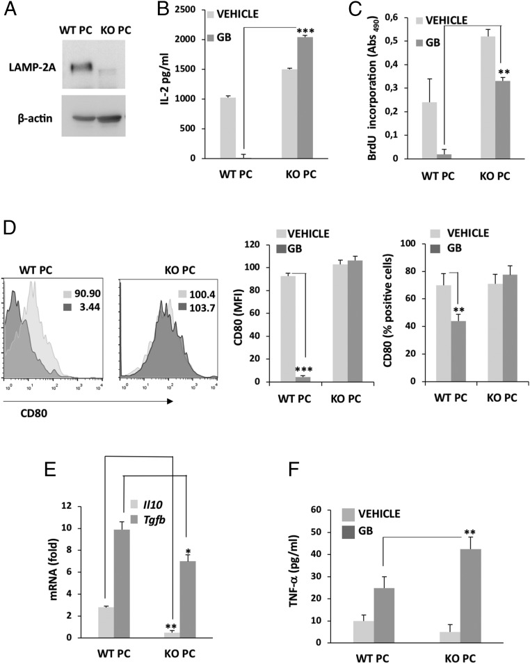
Fig. 2.
GB-induced CMA activity in PC is required for the acquisition of an immunosuppressive function in PC following interaction with GB. (A) Immunoblot of LAMP-2A in PC of LAMP-2A KO (KO PC) or WT mice (WT PC). (B) IL-2 production was measured by ELISA after 72 h of stimulation of naïve OT-II CD4+ T cells in response to OVA323–339 peptide presented by KO PC conditioned (GB) or not (vehicle) by GB and compared to control WT PC. Values are normalized to basal levels of IL-2 in resting T cells. Results are mean + SD from 5 to 6 different experiments; ***P < 0.001. (C) T cell proliferation was measured by BrdU incorporation in response to antigen presentation by WT PC and KO PC previously conditioned (GB) or not (vehicle) by GB. Results are mean + SD from 4 different experiments, **P < 0.01. (D) Flow cytometry analysis of the expression of the costimulatory molecule CD80. Insert numbers inside histograms represent MFI values. Bar graphs show MFI values and percentages of cells expressing CD80. Nonspecific fluorescence was measured using specific isotype monoclonal antibody and GB cells were used as negative control. Data represents mean ± SD obtained from at least 3 independent experiments, **P < 0.01; ***P < 0.001. (E) mRNA expression of Il10 and Tgfb assessed by qPCR in GB-conditioned WT PC and KO PC relative to basal levels in cells cultured in the absence of GB. Data (mean + SD from 3 different experiments) are presented as fold-induction of mRNA expression following coculture with GB for 72 h. *P < 0.05, **P < 0.01. (F) ELISA measuring TNF-α secreted by WT PC and KO PC cocultured with (GB) or without (vehicle) GB cells for 72 h. GB cells were used as negative control. All data represent mean ± SD obtained from at least 3 independent experiments; **P < 0.01.

