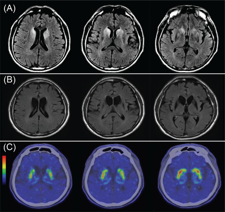
Figure 1.
Structural and Functional Imaging of the Patient’s Brain. Brain MRI nonenhanced T2 FLAIR images acquired 4 months (A) and 12 months (B) after initial symptom showed marked striatal hyperintensity and striatal atrophy, respectively, and FP-CIT PET scan showed a decrease in DAT binding in the bilateral striatum (C). Abbreviations: [18F] N-(3-fluoropropyl)-2β-carbomethoxy-3β-(4-iodophenyl) nortropane (FP-CIT) positron emission tomography (PET); DAT, Dopamine Active Transporter; FLAIR, Fluid Attenuated Inversion Recovery; MRI, Magnetic Resonance Imaging.

