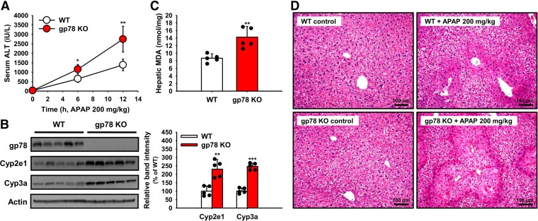
Fig. 10.
In vivo APAP-elicited liver injury in WT and gp78-KO mice. After an overnight fast (12 hours), mice (N = 5 in each group) were treated with APAP and their serum ALT levels determined at 0, 6, and 12 hours (A) as detailed (Materials and Methods). Mice were killed at 12 hours and their livers used for Cyp2e1 and Cyp3a IB analyses (B) and hepatic MDA levels (C) as described (Materials and Methods). The five individual values used for the MDA (mean ± S.D.) values are included in the bar graph. Statistical significance determined by the Student’s t test, *P < 0.05 or **P < 0.01 vs. WT. Liver injury upon hematoxylin and eosin staining and microscopic histologic analyses of representative control and APAP-treated WT and gp78-KO mouse livers isolated at 12 hours is shown in the right panels (D). ***P < 0.001 vs. WT.

