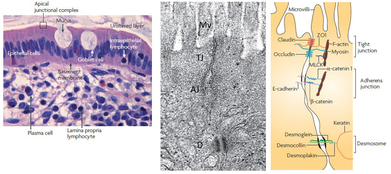
Figure 1: Anatomy of the mucosal barrier.
Left panel: In the human intestinal mucosa, composed of columnar epithelial cells, lamina propria (with its immune cells) and muscular mucosa, the goblet cells synthesize and release mucin, and the unstirred layer is immediately above the epithelial cells. The tight junction is a component of the apical junctional complex, and it seals paracellular spaces between epithelial cells.
Middle and Right Panels show an electron micrograph and the corresponding line drawing of the junctional complex of an intestinal epithelial cell. The key elements of the tight junction are the zona occludens and zona adherens, each of which is made up of different components. Just below the base of the microvilli (Mv), the plasma membranes of adjacent cells seem to fuse at the tight junction (TJ), where claudins, zonula occludens 1 (ZO1), occludin and F-actin interact. E-cadherin, α-catenin 1, β-catenin, catenin δ1 (also known as p120 catenin; not shown) and F-actin interact to form the adherens junction (AJ). Myosin light chain kinase (MLCK) is associated with the peri-junctional actomyosin ring. Desmosomes, located beneath the apical junctional complex, are formed by interactions between desmoglein, desmocollin, desmoplakin and keratin filaments.
In general, diffusion through claudins and occludin within the membrane is energy-independent, whereas ZO-1 facilitates exchange between tight junction and cytosolic pools through energy –dependent mechanisms
Reproduced from ref. 17.

