Figure 5.
Impaired Trafficking and Vacuolar Degradation of ABA Receptors in alix-1 Mutants.
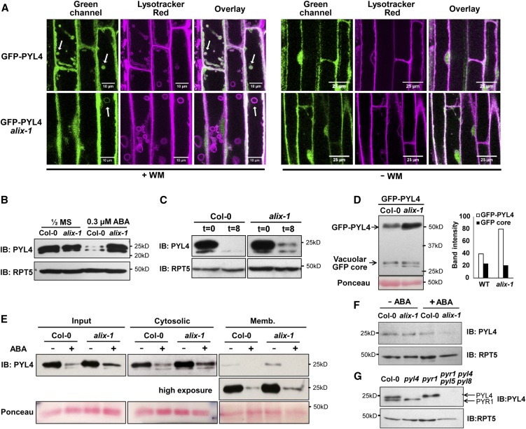
(A) Confocal images of wild-type (Col-0) and alix-1 mutant root cells expressing GFP-PYL4 on treatment or not with wortmannin (WM) and stained with LysoTracker Red. Bars = 10 and 25 µm. Arrows indicate representative WM-enlarged vesicles.
(B) Immunoblot (IB) showing accumulation of endogenous PYL4 in wild-type (Col-0) and alix-1 seedlings grown in the presence or absence of 0.3 µM ABAμ. Anti-RPT5 was used as loading control.
(C) Immunoblot (IB) analyses of PYL4 levels in 8-d-old wild-type (Col-0) and alix-1 seedlings treated with 50 µM CHX for 8 h. Anti-RPT5 was used as loading control.
(D) Immunoblot (IB) analysis to track the vacuolar delivery of GFP-PYL4 in the wild-type (WT, Col-0) and alix-1 backgrounds. Anti-GFP was used in immunoblots to detect GFP-PYL4 and the GFP-core signal. Band intensity was quantified using Fiji (ImageJ 1.52i). Ponceau staining was used as loading control as indicated.
(E) Isolation of microsomes from postnuclear fractions (Input) of 8-d-old wild-type (Col-0) or alix-1 seedlings treated or not with ABA for 3 h. Endogenous anti-PYL4 was used to evaluate PYL4 abundance in the cytosolic and microsomal (Memb.) fractions. Ponceau staining was used as loading control as indicated.
(F) Immunoblot (IB) analysis of PYL4 levels in nuclear protein extracts from 8-d-old wild-type (Col-0) or alix-1 seedlings treated or not with ABA for 3 h. Anti-RPT5 was used as loading control.
(G) Immunoblot to test the specificity of anti-PYL4. Protein extracts from 8-d-old wild-type (Col-0), single mutants pyr1 and pyl4 and quadruple mutant pyr1 pyl4 pyl5 pyl8 were used. Arrows indicate the position of endogenous PYR1 and PYL4 proteins in the immunoblot. Anti-RPT5 was used as loading control.

