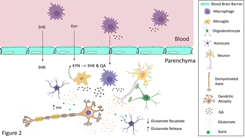
Figure 2: Heuristic Model of the Pathological Effects of Neurotoxic Kynurenines.
Under inflammatory conditions circulating macrophages produce more kynurenine (KYN), 3-hydroxykynurenine (3HK), and quinolinic acid (QA). 3HK can cross the blood brain barrier damaging neuronal cells through the production of free radicals. KYN also crosses the blood brain barrier where it is preferentially metabolized into 3HK and QA by microglia. Further, macrophages can infiltrate the brain parenchyma, where they are estimated to produce 32 times more QA than resident microglia182. Thus, although QA is usually found in low nanomolar concentrations in the human brain and CSF, a significant increase in QA levels to micromolar concentrations likely occurs in patients with neuroinflammation183. QA may contribute to the excitotoxic processes caused by the deficient glutamate reuptake (and paradoxical release) by dysfunctional astrocytes. Dendritic atrophy and remodeling also likely occurs altering functional connectivity. Further, 3HK and QA may damage oligodendrocytes leading to white matter abnormalities. Oligodendrocytes are highly sensitive to inflammation and reductions in their numbers or density are one of the most prominent findings in mood disorders at postmortem40. Note that other inflammatory mediators also play an important role in these neuropathological processes. Only the kynurenines are shown in the figure for clarity.

