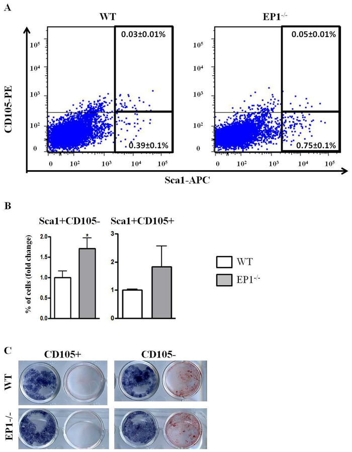
Figure 3: EP1−/− periosteum contains more CD105− progenitors.
A-B, freshly isolated periosteal cells were stained with antibodies against: CD45-PrcP, CD31-PEcy7, CD105-PE, Sca1-APC (A-B). The cells were selected for CD45 and CD31 negative cells and the percentage of Sca1 + and CD105+ cells were analyzed. (C), Sca1+CD105+ and Sca1+CD105− cells were sorted and cultured with osteogenic medium for 10 days. In the paired culture wells shown in the figure, the well on the left is stained for alkaline phosphatase and the well on the right is stained with alizarin red to detect mineralization in the cultures.
N=5 replicates per group for each assay. Error bars represent standard error of the mean. Statistical analysis was performed using paired student t-test. (*)=p<0.05 (**)=p<0.01 vs. age-matched WT.

