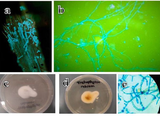
Figure 2.
(A) Endothrix hair invasion with green, fluorescent hyphae and Trichophyton violaceum spores (Calcoflour white stain, fluorescent microscope, ×400). (B) Fluorescent septate hyphae of Trichophyton rubrum against a darker background in a nail sample (Calcoflour white stain, fluorescent microscope, ×100). (C) Anterior surface of a Trichophyton rubrum culture. (D) Reverse surface of a Trichophyton rubrum culture. (E) Microscope image of blue, thin walled Trichophyton rubrum macroconidia showing the clavate shape and multiseptate, smooth walled structure (lacto phenol cotton blue stain, ×100).

