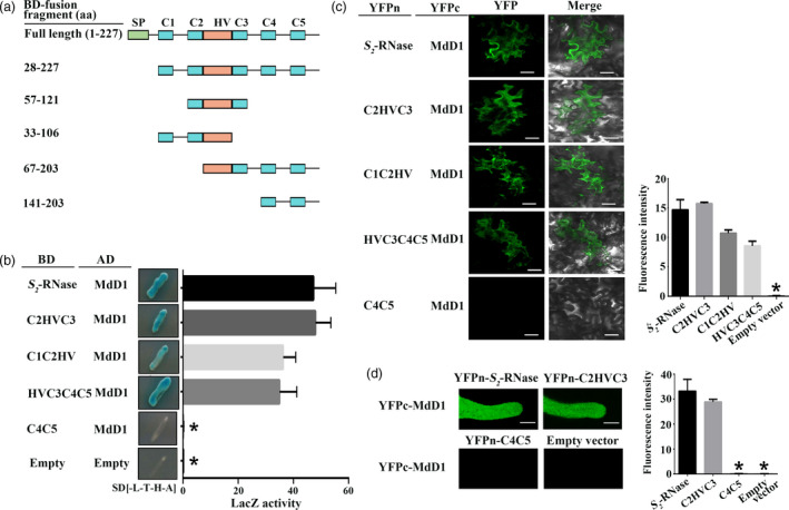
Figure 6.

MdD1 interacts with the RNase activity site of S‐RNase. (a) A segment model of S‐RNase. The sequence of S‐RNase was divided into four fragments according to the RNase activity site of S‐RNase. (b) Yeast two‐hybrid (Y2H) assay showing the interactions between MdD1 and different fragments of S 2 ‐RNase as described in (a). The active sites of S 2 ‐RNase are localized in the C2 and C3 domains, and S 2 ‐RNase was divided into four fragments. (c) Bimolecular fluorescence complementation (BiFC) assay showing the interactions between MdD1 and different fragments of the S 2 ‐RNase. Scale bars = 10 μm. (d) BiFC assay showing the interactions between MdD1 and different fragments of the S 2 ‐RNase in pollen tube. Scale bars = 10 μm.
