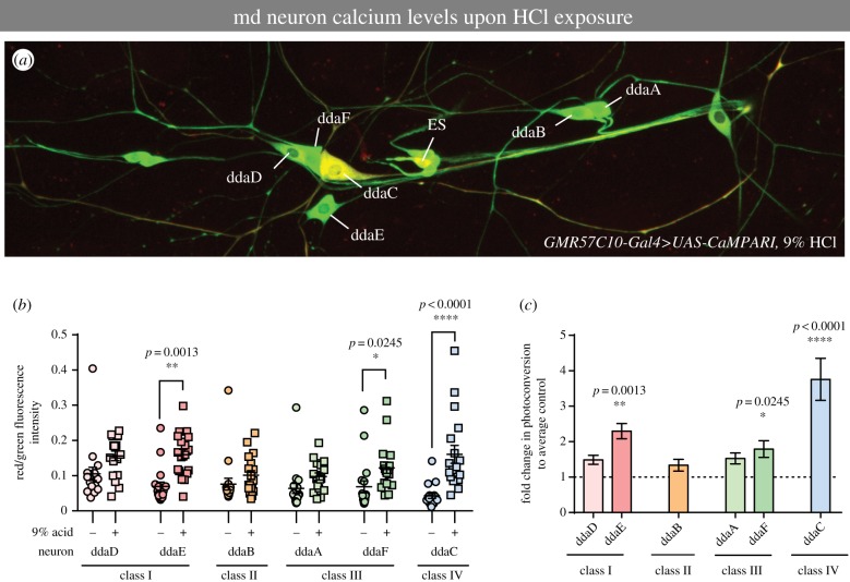
Figure 4.
Acid activates class IV nociceptive neurons. (a) Representative image of CaMPARI photoconversion in the larval dorsal cluster of md sensory neurons following treatment with 9% HCl and violet light exposure. The class IV ddaC neuron visibly exhibits the highest level of red fluorescence relative to other md neurons subclasses/subtypes (ddaA/B/D/E/F). External sensory neuron (ES) also displayed relatively high fluorescence levels. (b) Red/green CaMPARI fluorescence intensity quantification for each md neuron class/subtype in control mock-treated (no HCl) or 9% HCl-treated third instar larvae. Each circle/square represents an individual neuron of the specified class. (c) Fold difference in green-to-red CaMPARI photoconversion by md neuron class/subtype when compared with mean vehicle control. (b,c) Data are means ± s.e.; n = 18 neurons; asterisks (*) indicates significant difference in CaMPARI photoconversion when compared with mock-treated vehicle control. Statistical significance between vehicle control and acid groups was determined via Kruskal–Wallis with Dunn's multiple comparisons test.

