Figure 7.
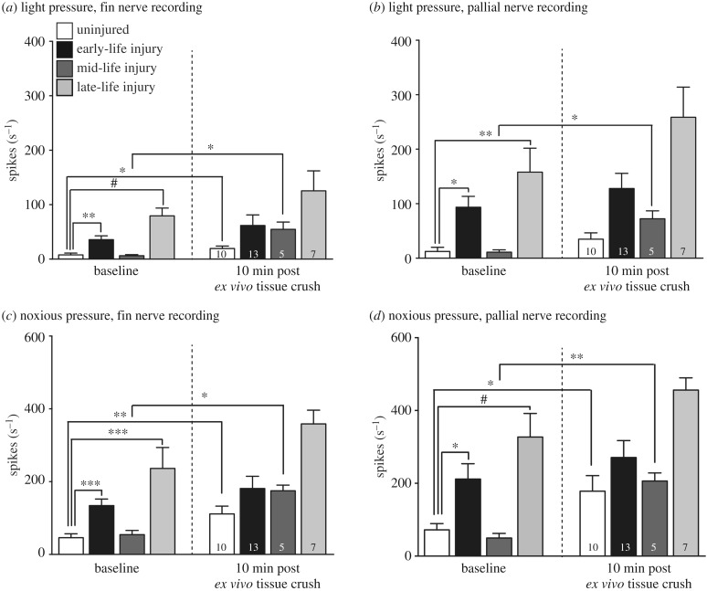
Paired fin/pallial electrophysiological recordings of adult squid. (a) Injury in early life produces permanent hyperexcitability in the fin nerve when the fin tissue in stimulated with a light von Frey filament, compared with uninjured squid. This pattern is not present in MLI squid. Squid injured acutely (24 h prior to recording) in late life also show hyperexcitability compared with controls. In tests of short-term, ex vivo sensitization in response to tissue crush, there was an increase in spikes produced in squid that did not receive an in vivo injury (uninjured), and in squid injured in mid-life. No plasticity was evident in the ELI or LLI groups. (b) A similar pattern is apparent in the pallial nerve when fin tissue was stimulated with a light von Frey filament. In general, early-life and LLI groups showed sensitization at baseline recording, while short-term plasticity occurred only in the MLI group. (c) When the fin tissue was stimulated with a heavy von Frey filament producing noxious pressure, the same pattern of long-term and short-term plasticity was seen as in (a). (d) In response to noxious pressure on the fin, pallial nerve excitability was the same as for (b), except that short-term plasticity is also apparent in the uninjured group. Mixed model ANOVA followed by post hoc, sequential Bonferroni-corrected t-tests. Comparisons among groups at baseline made with unpaired t-tests. Comparisons within groups before and after ex vivo tissue crush made with paired t-tests. *p < 0.05, **p < 0.01, ***p < 0.001, #p < 0.0001. Numbers inside bars are group sample sizes (same for baseline and post-crush). Bars show mean + 1 s.e.m.

