
Figure 3.
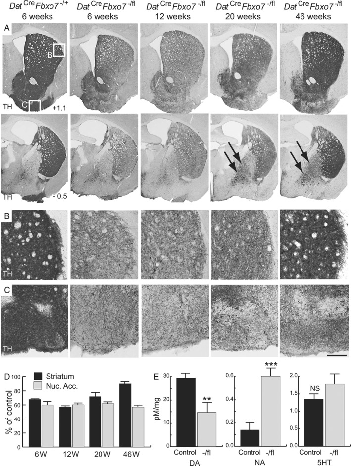
Decreased TH+ staining in mice with a conditional loss of Fbxo7 expression in dopaminergic cells. (A) TH‐immunostained sections of the striatum at +1.1 mm (top panels) and −0.5 mm (bottom panels) relative to the bregma from control mice (Dat Cre Fbxo7 −/+) at 6 weeks, and conditional KO mice (Dat Cre Fbxo7 −/fl) at 6, 12, 20, and 46 weeks of age. Arrows show increased TH staining in the globus pallidus over time. (B, C) Magnified regions highlighted in A, showing TH+ fibres in the striatum (B) and olfactory tubercle (C). (D) Quantification of TH+ fibre density in Dat Cre Fbxo7 −/fl mice at different time points, as a percentage of control at each time point, in the striatum and nucleus accumbens (Nuc. Acc.). (E) Quantification of neurotransmitter levels in the striatum of 5‐week‐old Dat Cre Fbxo7 −/+ and Dat Cre Fbxo7 fl/+ mice (control) and Dat Cre Fbxo7 −/fl mice (−/fl) using HPLC analysis. DA, dopamine; NA, noradrenaline; 5HT, serotonin. **p < 0.01; ***p < 0.001. NS, not significant. The scale bar in C represents 800 μm for A and 300 μm for B, C.