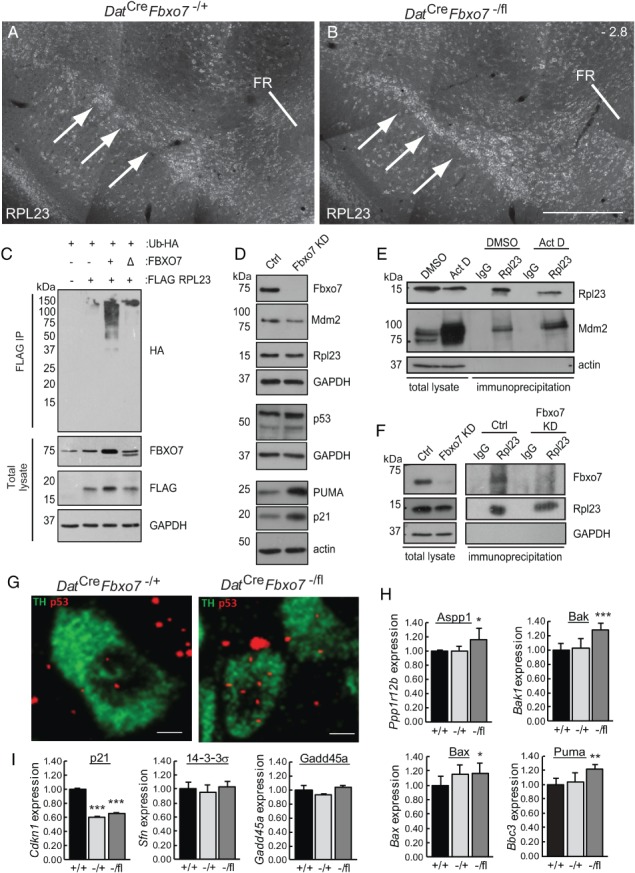
Figure 6.

Mutant mice show increased RPL23 and elevated expression of Trp53 mRNA and p53‐regulated pro‐apoptotic genes. (A, B) Immunofluorescence for RPL23 (arrows) in the SNpc of control (A) and Dat Cre Fbxo7 −/fl (B) mice at bregma stage −2.8. FR, fasciculus retroflexus (n = 3). The scale bar in B represents 200 μm for A, B. (C) In vivo ubiquitination assay of RPL23 using HEK293T cells transfected with plasmids expressing FLAG‐RPL23, HA‐tagged ubiquitin, and either untagged WT (+) or ΔF‐box (Δ) FBXO7 constructs. Immunoblots of total lysate prior to anti‐FLAG immunoprecipitation are also shown. (D) Immunoblotting for the expression of various proteins as indicated from cell lysates of SHSY‐5Y cells either constitutively expressing an shRNA targeting Fbxo7 expression or a control. (E) Co‐immunoprecipitation assays from SHSY‐5Y cells treated for 8.5 h with 5 nm actinomycin D or vehicle (DMSO). Lysates were immunoprecipitated with antibodies to RPL23 and immunoblotted for MDM2. (F) Co‐immunoprecipitation assays from SHSY‐5Y cells with endogenous (Ctrl) or reduced expression of FBXO7. Cells were treated for 4 h with 5 nm actinomycin D and then for a further 5 h with MG132 prior to harvesting. Cell lysates were immunoprecipitated with RPL23 antibodies and immunoblotted with antibodies to Fbxo7. (G) Th (green) and Trp53 (red) transcripts were labelled using branched DNA amplification in situ hybridisation. (H, I) RT‐qPCR analysis of p53‐regulated genes isolated from dissected midbrains isolated from Dat Cre Fbxo7 +/+ (+/+), Dat Cre Fbxo7 −/+ (−/+), and Dat Cre Fbxo7 −/fl (−/fl) mice. Expression was normalised to three reference genes (Actb, Ppia, Gapdh) and expressed relative to WT levels. *p < 0.05, **p < 0.01, ***p < 0.001.
