
Figure 2.
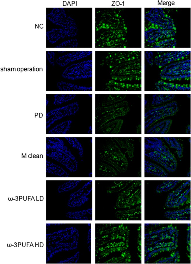
Immunofluoresence of ZO‐1 expression in the ileum. Original magnification, 400×. Changes in spatial expression of ZO‐1 protein were qualitative only. Same slide was stained with rabbit anti‐ZO‐1 antibody, followed by Alexa fluor 488 conjugated IgG (ZO‐1). Nuclei were further stained with DAPI (DAPI). The pictures of ZO‐1 and DAPI were merged (Merge). DAPI, 4′,6‐diamidino‐2‐phenylinodele; PD, peritoneal dialysis; ω‐3PUFA LD, ω‐3PUFA low dose; ω‐3PUFA HD, ω‐3PUFA high dose.