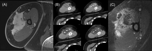
Figure 1.

(A) Pre‐ablation axial T1 postcontrast MRI and (B) procedural images obtained for a patient undergoing cryoablation of an upper extremity desmoid tumor. Sequential axial noncontrast CT images obtained intermittently throughout the procedure reveal a progressive increase in the ablation zone that ultimately encompasses the mass. (C) First follow‐up axial T1 (fat sat.) postcontrast MRI revealing a small area of residual enhancing tumor at the posterior ablation margin, which was treated with a second cryoablation procedure, with extensive heterogenous enhancement anteriorly consistent with expected posttreatment change. MRI, magnetic resonance imaging
